Introduction
In radiotherapy, surface dosimetry is very complicated
issue. The reason of this complication is the rapid dose
change between the dose entrance point and the maximum dose depth. Compton interactions are usually the
main reason of radiation absorption in tissues. When
the primary photon beams interact with a homogenous
material, secondary particles such as electrons or
positrons are liberated and by the increasing the depth into the material up to a point (maximum depth dose),
more secondary electrons are set in motion by secondary
particles. Due to the having finite path lengths,
number of electrons reaches a maximum at a particular
depth and begin to decrease because the beam is attenuated
as the depth increases. At this particular depth,
the number of generated electrons is equal with the
number of electrons which are stopped and deposited
their energy. It is said that the charged particle equilibrium
(CPE) exists at this point. In the near surface region,
the required conditions of CPE are not achieved
and even the dose gradient is quite high. Lack of CPE
and being a high dose gradient region make the surface
dosimetry complicated.[]
The dose determination of a problematic region
such as buildup region, specific dosimetric considerations
should be taken. In treatment planning systems
(TPSs), to get the accurate dose calculation depends
on the selection of appropriate dosimetric systems and
their correct use in dose measurements. For the precise
beam modeling of the algorithms inside TPS, dose measurements
required for modeling should be done with
great precision. Especially in the problematic region as
skin surface and buildup region, to avoid skin damage,
to decide the thickness of the bolus material and radiotherapy
treatment technique or the dose fractionation
scheme, the amount of surface dose calculated by TPS
should be known correctly. On the other hand, the dose
calculation accuracy on the skin surface is a rather controversial
issue. Unless they are well-modeled, the most
commercial TPSs usually cannot calculate the surface
dose accurately.[] After a certain depth, the accuracy
of the TPS can be considered satisfactory. In general,
TPS algorithms tend to over or under-estimate surface
dose. For example, Higgins et al.[] reported that at the
near surface, dose deviation between the calculated with
Eclipse TPS and measured with 1 mm3 thermoluminescent
dosimeters (TLD) chips is between -12% and 10%.
In the anthropomorphic phantom study of Qi et al.,[]
it was reported that the Peacock system calculates the
surface doses up to 8.5% higher than the measuring systems.
Panettieri et al.[] have investigated the calculation
accuracy of Anisotropic Analytical Algorithm (AAA)
and PBC algorithms between the surface and 2 cm depth
and they compared the calculated data with Monte Carlo
simulation. They showed that both algorithms underestimate
the absorbed dose in the build-up region.
The selection of dosimetry systems for buildup region
dose measurement is very vital. For a precise beam
modeling in TPS, surface dose should be measured
within at least ±5% accuracy. Extrapolation chambers, Attix chambers, TLDs, film dosimeters, and metal oxide
semiconductor field effect transistors are the most
common dosimetry systems used in surface dosimetry.
For a more reliable dose measurement in buildup region,
extrapolation chambers are suggested but these
tools can be accessed only by a few institutions. Parallelplate
ion chambers are good alternatives to the extrapolation
chambers in surface dosimetry, but due to their
over respond behaviors in high dose gradient region,
some correction factors should be used (Gerbi and
Khan's method).[] When the appropriate corrections
for polarity, obliquity, and in-scattering are applied to
parallel-plate chamber measurements, similar results
can be obtained to the extrapolation chambers considered
as gold standard in the surface dose.[] According
to the study of Zhuang and Olch[] the results of the
Markus parallel plan ion chamber are compatible with
the Monte Carlo simulation in the build-up region. Absolute
dose differences were found within the 3%.
Films are one of the passive detectors used in surface
dosimetry. Gafchromic films are commonly used
in radiotherapy dosimetry today because of their practical
usage. They are insensitive to light, independent
from dose fraction, dose rate and energy, do not require
bathing as in radiographic films, and have a high spatial
resolution. Because of their water equivalent feature,
they are frequently preferred in in vivo dosimetry (IVD).
IVD is one the quality control method in radiotherapy.
This method allows the measurement of the dose
that reaches the patient during treatment and therefore
it can be seen as the most reliable quality control
method in radiotherapy. The dose control of the treatment
is achieved by placing the detectors in natural
body cavities or on the patient skin. Gafchromic films
can be cut into small pieces,[] give the users two-dimensional
map of surface dose, and can record radiation
permanently. Due to these favorable features, it is
frequently preferred in IVD applications for skin dose
measurements. Cao et al.[] reported that superficial
dose measurements obtained by Monte Carlo simulation
are compatible with multilayer Gafchromic film
results. In superficial region, they show a good agreement
by the mean difference <1%.
The uncertainties and error probabilities that may
arise in the progress of radiotherapy techniques can
also increase. Despite the possible uncertainties that
may derive from the characteristics of the modern
radiotherapy treatment techniques, determining the
dose applied to the target volume will be useful in controlling
the accuracy of these techniques.[] The comparison
of the surface doses calculated in the TPSs and measured with different dosimetric systems is one of
the techniques in the quality control programs of treatment
plans. For this reason, surface dose responses of
TPSs used in clinics and dosimetric systems used in patient
surface dosimetry should be controlled by surface
dosimetry systems which are accepted as reference.
In this study, the calculation accuracy of Acuros
XB (AXB) and AAA v15.1algorithms is investigated
and compared at the buildup region with parallel-plate
ionization chamber and EBT3 films in different field
sizes for 6 MV photon energy. The behavior of the
Gafchromic EBT3 film in surface region was checked
by Markus parallel-plate ion chamber and after that
the calculated results were compared with film. It was
aimed that to determine the surface dose differences of
the calculated by different algorithms and measured by
film and to consider it during the quality control of the
patient plans with IVD.
Methods
Phantom and Parallel-Plate Ion Chamber Dose Measurements
Varian Trilogy linear accelerator (Varian, Palo Alto, CA)
equipped with a Millennium 120-leaf MLC was used in
this study. All near surface central axis depth dose measurements
were performed on the 6-MV photon beam
along the central axis with open field sizes of 5×5, 10×10,
and 20×20 cm2 at a constant source-to-surface distance
of 100 cm. Since the measurements to be taken include
problematic dose regions, SSD has not been changed
to provide the necessary sensitivity for distance when
solid water phantom is added on the plate to be taken
at different depths. Then, source to skin distance correction
for SSD=100 cm was applied to the results for each
dosimeter. The measurements were performed in 40×40
cm2 solid water equivalent phantom slabs (SP34, PTW
Freiburg, Freiburg, Germany) and the slab thickness
varied from 1 to 10 mm. The physical density of solid
water phantom is 1.045 g/cm-3. We obtained the central
axis depth dose in the near surface region using the
parallel-plate ionization chamber (Markus 23392, PTWFreiburg)
and the EBT3 Gafchromic film (International
Specialty Product, NJ, US). To acquire the average values,
measurements were repeated 3 times.
The penetration range of the ion beam in a material
is usually characterized by water equivalent thickness
(WET). WET measures the amount of liquid
water thickness that can stop the ion beam, as does a
material of a certain thickness.[] WET is calculated by
taking into account the density of RW3 water equivalent phantom and the effective measurement depths of
each dosimeter systems. If the dose responses of different
dosimetry systems are to be compared at the same
depth, the WET values must be known. Interpolation or
extrapolation methods can also be used to obtain doses
for different dosimeters at specific depths. WET values
were calculated according to the effective measurement
points of the dosimeters and hereby, depths corresponding
to the read doses were obtained for all dosimetry
systems. Interpolation and extrapolation methods were
used to find the dose values at the same depths.
The recommendation of Commission on Radiation
Units and Measurements and the International Commission
on Radiological Protection about skin depth
for surface dosimetry is 0.07 mm8 since this depth is
generally corresponds to the interface between the dermis
and epidermis layers of the skin. In our study, the
comparisons were made at this recommended depth
and 1, 2, 3, 4, and 5 mm which are accepted as near
surface region.
In our study, one of the dosimeters used in the surface
dose measurements is Markus parallel-plate ion
chamber. The effective measurement point of Markus
ion chamber is 0.023 mm which corresponds to the inner
surface of the proximal collecting plate. The central
axis near surface depth dose measurements were performed
for 6 MV photon beams, at a phantom depth
of 0, 1, 2, 3, 4, and 5 mm and 100 cm fixed SSD. Three
readings were taken using the Unidos Webline electrometer
(PTW Freiburg, Germany). The readings were
taken for both positive and negative voltage (±300 V)
to take into account the polarity effects. Hereby the polarity
correction factor was found and applies to readings.
SSD correction factors were applied to the results.
100 MU was delivered for each measurement and the
results were normalized to 15 mm which is accepted as
the maximum dose depth of 6 MV photon beams.
Some electrons scattered from the side walls of
fixed-separation parallel-plate ion chamber and in the
buildup region they mainly contribute to the charge
and these results to get over response dose by parallelplate
ion chamber. Velkley et al.,[] first introduced
a correction factor for this perturbation effect. Gerbi
and Khan improved the previous formula by including
some parameters about parallel-plane ion chamber.
The correction equation of GK (Gerbi and Khan)
method[6] was given by Eqs. (1) and (2) below:
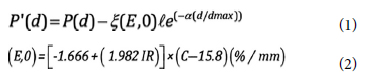
Where P (d) is corrected and P"(d) is uncorrected
percentage depth dose at "d" depth. (E,0) is the energy
dependent chamber factor that indicates the over response
in percent per mm of chamber plate separation
at the phantom surface; ? is the plate separation and
it is 2 mm for PTW Markus ion chamber; d and dmax
represent the depth of the chamber front window and
maximum dose depth respectively. IR is the ionization
ratio that measured at 10 and 20 cm depths, 10×10 cm2
field size at SSD 100 cm. For 6 MV photon beam, IR
is 0.6709. C is the distance between the collector edge
and side wall, and it is regarded as 0.35 mm for PTW
Markus ion chamber. α is a constant and equal to 5.5.
All factors were placed in equations above and the over
response correction factors were obtained. Thus, Grebi
and Khan's overdose correction method[6] was applied
to our PDDs.
Film Dose Measurements
In the present study, we used Gafchromic EBT3 films
(International Specialty Product, NJ, US) from the
same batch which has a single active layer of approximately
30 ?m thickness. Gafchromic EBT3 film has a
30 ?m thickness of single active layer which is between
125 ?m thick of transparent polyester sheets. The effective
measurement point of EBT3 film was assumed
at the depth of 0.153 mm.[] A calibration curve was
created to accurately evaluate the irradiated films and
the irradiated films were calibrated according to this
curve. To generate film calibration curve, films were
cut into small pieces. They were placed at 5 cm depth
which was defined as the depth of calibration for 6 MV
photon beams. All films were irradiated within the
dose ranges of 0-800 cGy at the field of 10×10 cm2. One
of the films was released for the background without irradiation.
After 24 h, all the films were scanned by Epson
Expression 10000XL scanner. Irradiated films were
analyzed in Image J program and separated into three
different channels (red, blue, and green channels). In
Gafchromic films, the largest contrast difference is seen
in red channel therefore red color was selected for dose
reading. The film calibration program in Mephysto
mcc software (PTW Mephysto mc2, PTW, Freiburg,
Germany) was used to create calibration curve. Using
this program, the optical densities in the center of
scanned films was read. A calibration curve was created
based on the permeability values corresponding
to the dose values.
Film measurements were performed at the same
setup conditions as the ion chamber measurements.
Due to the different effective measurement point of film, the WET values corresponding to the measured
doses were calculated carefully.
TPS Dose Calculations
Varian Eclipse TPS (Varian Headquarters, Palo Alto,
California, USA) which has AAA 15.1 versions and AXB
algorithms was used for TPS dose calculations. In TPS,
a water phantom size of 40×40×10 cm3 was formed automatically
and beams were set on it in the same field
sizes and gantry angles as described in parallel-plate
ion chamber and Gafchromic film measurements. The
fraction dose for each plan was set at 100 MU, the doses
which the dosimetry systems were irradiated also. All
the plans were calculated with different algorithms for
the same setup conditions. The calculation grid size of 1
mm was used for all calculation algorithms. Doses were
read at the depth of effective measurement of film and
ion chamber from the TPS. The calculated PDDs were
normalized the dose at 1.5 cm depth for each field sizes.
Results
The doses in the near surface for 6 MV photon beams
were measured with Gafchromic EBT3 film and
Markus parallel-plate ion chamber for the square field
sizes of 5×5, 10×10 and 20×20 cm2. The effective measurement
point of both dosimetry systems was noted
and to obtain doses at 0.07, 1, 2, 3, 4, and 5-mm depth
for each dosimetry, interpolation, and extrapolation
were made using four-order polynomial functions. The
percentage doses at the surface and buildup regions for
different field sizes measured using the Markus parallel
plane ion chamber are given in Table 1.
Table 1: Percentage depth doses (PDDs) obtained with
a Markus parallel plane ion chamber in a water
equivalent RW3 phantom using 6MV photon
beams for different field sizes at SSD=100 cm.
The interpolated and extrapolated skin dose values
at 0.07 mm for Markus ion chamber and EBT3 film are
14% and 12.79% for 5×5 cm2; and 30.87% and 27.07%
for 20×20 cm2, respectively. The PDD curve comparison
for the measurement for a 10×10 cm2 field size is
shown in Figure 1.
Fig 1: Percentage depth doses (PDDs) at surface and
buildup regions for 6 MV photon beams using a
Markus parallel plate ion chamber and Gafchromic
EBT3 film.
PDD: Percentage depth dose, WET: Water equivalent
thickness.
Markus parallel plane ion chamber can be considered
as a reference dosimetry in surface dosimetry. On
the other hand, because of their physical characteristics
they cannot be used in IVD. Its characteristics such as
low spectral sensitivity and high spatial resolution make
the film an appropriate dosimeter system in surface and
buildup region dosimetry. In Figure 1, it is seen that in
the near surface EBT3 film shows close results with the
Markus ion chamber for all field sizes and depths.
Each dosimeter has a different effective measurement
depth. To obtain percentage depth dose values
of film and TPS at the same depths, WET of the systems
should be considered. The WET values belong to
Gafchromic EBT 3 Film are given in Table 1 and they
were used to obtain the PDD values from TPS for the
same depths. The PDD results at 0.07 mm for 5×5,
10×10, and 20×20 cm2 field sizes acquired by TPS algorithms
and Film are shown in Figure 2.
Fig 2: Percentage depth doses varying with field sizes at
0.07 mm which is the recommended by ICRU as
surface dose depth.
PDD: Percentage depth dose, AAA: Anisotropic Analytical
Algorithm.
As shown from Figure 2, for all field sizes AAA
v15.1 shows highest results. For 10×10 and 20×20 cm2,
surface depth doses calculated by Acuros algorithm
and measured by Gafchromic EBT 3 film are nearly
similar. On the other hand, as shown in Figure 3, for
all field sizes by increasing depth, all algorithms and
film depth dose values were approaching to each other.
Fig 3: Percentage depth doses (PDDs) for 6MV photon
beams with different sizes at 100 cm fixed SSD.
The doses at 0, 1, 2, 3, 4, and 5mm depths were
obtained by Gafchromic EBT 3 film and Eclipse
TPS with AAA v15.1 and Acuros algorithms, and
the following field sizes were investigated: 5×5
cm2 (a), 10×10 cm2 (b), and 20×20 cm2 (c).
PDD: Percentage depth dose, AAA: Anisotropic Analytical
Algorithm, WET: Water equivalent thickness.
Discussion
Although the surface dose measurement is a challenging
issue, it is the best reliable QA method to control
the dose that reaches the patient during treatment
which is called IVD. One of the most preferable IVD
methods is to control of the skin dose by placing the
dosimeter on the patient skin. By this method, the
quality of the patient treatment plans can be checked
in the most correct way.
To measure doses of buildup region accurately,
dosimetry preference is very crucial. As known from
previous studies, extrapolation chambers are the recommended
dosimeters for surface dosimetry.[] Nevertheless,
Markus parallel plane ion chamber can be a
good alternative to extrapolation chambers. Although
Markus parallel plate ion chambers are preferred for surface
dose measurements, they are not suitable for IVD
due to their physical properties. Therefore, in this study, it was only used to determine the accuracy of the film
in surface dose measurements as a reference dosimetry.
Radiochromic films are good options for surface
and buildup region dose measurements and are
suitable for IVD. In our study, Markus parallel plate
ion chamber was used as a reference dosimetry and
buildup region depth dose values were obtained.
The results were compared by Gafchromic EBT3
films. For 10×10 cm2 field size, surface doses at 0.07
mm with the Markus parallel-plate ion chamber and
Gafchromic EBT3 film were found 16.61% and 18.8%,
respectively. Close results show that also film can
be used as a reference dosimetry system for surface
dosimetry and it was used to control the accuracy of
TPS algorithms in our study.
The harder it is to measure the skin dose correctly,
the harder it is to calculate it through TPS. Algorithms
inside TPS cannot calculate buildup region doses correctly.
In our study, the film measurements were compared
by TPS results, calculated and measured surface
dose differences were obtained. These differences obtained
through the phantom will guide us while checking
the patient treatment plans through IVD.
Bilge et al.[] measured the surface dose with the
Markus parallel plate ion chamber for 6 MV (Siemens
Oncor Impression Plus linear accelerator) photon
beams and the result was found to be 15.0% for
a field size of 10×10 cm2. They also used EBT model
radiochromic film and found the surface dose at the
depth of 0 mm 20.0±2% for a field size of 10×10 cm2.
The surface dose with Gafchromic EBT3 film was
found to be 20.4% at the same depth and field size.
Qi et al.[] investigated the dose at the WET of
Gafchromic EBT3 film 0.153 mm which equals the
depth of 0 mm in the phantom, for the same field size
and the result was found 23.5%. Our study is consistent
with the previous studies.
Cao et al.[] investigate the superficial dose accuracy
of different algorithms comparing by Monte Carlo
algorithm and film measurements. Surface and buildup
region doses were measured by the group. They showed
that AAA algorithm overestimate skin dose near 4.07%
when compared by Monte Carlo simulation. Nevertheless
within 2-15 mm depth, results were compatible by
Monte Carlo.
Cho et al.[] studied about skin dose measurements
for lung stereotactic body radiotherapy. Measurements
were taken by GafChromic EBT2 film, Attix
chamber and compared to near surface skin doses
predicted by Eclipse TPS using the AAA algorithm for
a 6 MV photon beam. For 10×10 cm2 field size, surface
doses at 0 mm with the Attix chamber, GafChromic
EBT2, and AAA algorithm were found 16%, 23.9%,
and 40%, respectively. Their results show that AAA
algorithm overestimates the surface dose at the top of
the phantom (nominal 0 mm depth) compared to the
measured data by Attix chamber and EBT2 film.
In our study, the calculated results obtained by
AAA v15.1 and Acuros algorithms were compared by
Gafchromic EBT3 film. For 10×10 cm2 field size, surface
doses at 0.07 mm with the AAA v15.1, Acuros
algorithms and Gafchromic EBT3 were found 26.25%,
19.5%, and 18.8%, respectively. It was seen that for all
field sizes AAA v15.1 result was much higher than
film and Acuros algorithm. In addition to that, surface
doses obtained by Markus chamber and Gafchromic
EBT3 film at 0 mm depth were 16.61% and 20.40%
for 10×10 cm2 field size. The results present coherency
with the study of Cho et al.[]
According to the many researches, AXB algorithm
can calculate the dose accurately in regions with complex
geometries and heterogeneities.[,] The studies
about AXB algorithm were usually to investigate the
calculation capability of the algorithm in inhomogeneous mediums. There is not enough study about their
behaviors in the surface and buildup region. Alhakeem
et al.[] studied about the performance of different
dosimetric methods in inhomogeneous phantoms.
They used two versions of AXB and AAA algorithms,
EBT2 film and MOSkin dosimeters and compared the
results by Monte Carlo calculations. It was seen that
the newer version of AXB (v11.0.31) algorithm and
film results were closer to MC calculation than AAA
algorithm. In the near surface, AXB (v11.31) and
Gafchromic EBT2 results were seen similar. From the
near surface to 5 mm, Gafchromic EBT2 film showed
lower results than all algorithms; however, after 5 mm
all the values were approaching each other. In our results,
for all field sizes, in the buildup region, the results
obtained by AAA were found higher than AXB and
Gafchromic EBT3 results while AXB and Gafchromic
EBT3 results were close to each other. After 2 mm
depth, all the calculated and measured depth doses
were close to each other.
Conclusion
The accuracy of algorithms used to prepare the treatment
plans is an important issue. TPS has difficulties
in calculating, such as in heterogeneous or buildup regions.
Especially in the treatment of superficial cancers
or in the situation which the surface dose information
is crucial, the behaviors of algorithms in the buildup
region must be known precisely.
In clinics, to be sure of the correctness of surface
and buildup region dose calculation, QA procedures
must be performed for all algorithms implanted in TPS
before using them for patient treatment planning. By
this means, especially for treatment plans where surface
dose information is important, the behavior of the
algorithms in the surface region can be predicted. It
will be valuable to know the calculation capabilities of
the algorithms in the surface region in the quality control
of patient treatment plans with IVD.
Gafchromic EBT3 film is a useful dosimetry system
for IVD. Since it exhibits the similar results by Markus
parallel plate ion chamber in the surface and buildup
region, Gafchromic EBT3 film can be a preferable
dosimeter in QA programs for skin dose control of the
patients.
Peer-review: Externally peer-reviewed.
Conflict of Interest: All authors declared no conflict of interest.
Ethics Committee Approval: The study was approved
by the Academic Coordination Community of Istanbul
University Institute of Oncology Ethics Committee (No:
B.30.2.İST.0.53.00.00/911, Date: 28/03/2013).
Financial Support: None declared.
Authorship contributions: Concept - N.D.K., C.K.A.; Design
- N.D.K.; Supervision - N.D.K.; Funding - None; Materials
- N.D.K., C.K.A.; Data collection and/or processing -
N.D.K., C.K.A.; Data analysis and/or interpretation - N.D.K.,
C.K.A.; Literature search - N.D.K.; Writing - N.D.K.; Critical
review - N.D.K., C.K.A.
References
de Vera P, Abril I, Garcia-Molina R. Water equivalent
properties of materials commonly used in proton
dosimetry. Appl Radiat Isot 2014;83(Pt B):122-7.
Zhuang AH, Olch AJ. Validation of OSLD and a treatment
planning system for surface dose determination
in IMRT treatments. Med Phys 2014;41(8):081720.
Higgins PD, Han EY, Yuan JL, Hui S, Lee CK. Evaluation
of surface and superficial dose for head and neck
treatments using conventional or intensity-modulated
techniques. Phys Med Bio 2007;52(4):1135-46.
Qi ZY, Deng XW, Huang SM, Zhang L, He ZC, Li XA,
et al. In vivo verification of superficial dose for head
and neck treatments using intensity-modulated techniques.
Med Phys 2009;36(1):59-70.
Panettieri V, Barsoum P, Westermark M, Brualla L, Lax
I. AAA and PBC calculation accuracy in the surface
build-up region in tangential beam treatments. Phantom
and breast case study with the Monte Carlo code
PENELOPE. Radiother Oncol 2009;93(1):94-101.
Gerbi BJ, Khan FM. Measurement of dose in the
buildup region using fixed-separation plane-parallel
ionization chambers. Med Phys 1990;17(1):17-26.
Tatsiana AR, Patrick H. Surface dose measurements
with commonly used detectors: A consistent thickness
correction. J Appl Clin Med Phys 2015;16(5):358-66.
Devic S, Seuntjens J, Abdel-Rahman W, Evans M,
Olivares, M, Podgorsak EB, et al. Accurate skin dose
measurement using radiochromic film in clinical applications.
Med Phys 2006;33(4):1116-24.
Cao Y, Yang X, Yang Z, Qiu X, Lv Z, Lei M, et al. Superficial
dose evaluation of four dose calculation algorithms.
Radiat Phys Chem 2017;137(1):23-8.
Scarantino CH, Prestidge BR, Anscher MS, Ferree
CR, Kearns WT, et al. The observed variance between
predicted and measured radiation dose in breast and
prostate patients utilizing an in vivo dosimeter. Int J
Radiat Oncol Biol Phys 2008;72(2):597-604.
Velkley DE, Manson DJ, Purdy JA, Oliver GD Jr. Build-up region of megavoltage photon radiation sources.
Med Phys 1975;2(1):14-9.
Van Battum LJ, Hoffmans D, Piersma H, Heukelom S.
Accurate dosimetry with GafChromic EBT film of a 6
MV photon beam in water: What level is achievable?
Med Phys 2008;35(2):704-16.
Bilge H, Çakır A, Okutan M, Acar H. Surface dose
measurements with GafChromic EBT film for 6 and
18MV photon beams. Phys Med 2009;25(2):101-4.
Gwi AC, Ralston A, Mo TM, Martin D, Pickard S, Kim
JH, et al. In vivo and phantom measurements versus
Eclipse TPS prediction of near surface dose for SBRT treatments. J Phys 2014;489:012008.
Bush K, Gagne IM, Zavgorodni S, Ansbacher W,
Beckham W. Dosimetric validation of Acuros XB with
Monte Carlo methods for photon dose calculations.
Med Phys 2011;38(4):2208-21.
Lloyd S, Ansbacher W. Evaluation of an analytic linear
Boltzmann transport equation solver for high-density
inhomogeneities. Med Phys 2013;40(1):011707.
Alhakeem EA, Al Shaikh S, Rosenfeld AB, Zavgorodni
SF. Comparative evaluation of modern dosimetry
techniques near low and high-density heterogeneities.
J Appl Clin Med Phys 2015;16(5):142-58.



