

METHODS
This study included 60 post-operated left breast cancer patients who received chest wall/breast irradiation
by three-dimensional conformal radiotherapy (RT). The patients had received a total dose of 40Gy
in 15 fractions to whole breast/chest wall (two fields) and dose to supraclavicular fossa (three fields). We
compared the DVH for heart, lung, and spinal cord in both fields.
RESULTS
Mean dose for lung was 17.49Gy and for heart 12.31Gy, and spinal cord maximum dose was 0.78Gy.
The average lung NTCP for RP was 3.11%. The risk of RP was significantly greater in three-field radiation
as compared to two field for lung (p<0.05). We observed the statistically significant correlation
of MHD and NTCP (p<0.00001 and p=0.000402). The correlation of central lung distance (CLD) was
observed to be significant with three fields, that is, as the CLD increased risk of organ damage to lungs
increased (p=0.00022). The correlation of CLD and MHD with DVH was observed to be statistically
significant (p<0.001).
CONCLUSION
This study results are useful to reanalyze the previous 2D-based clinical reports about breast RT complications
as a view point of the NTCP. Further studies are needed for the actual clinical data of complications
with this DVH analysis.
Keywords: Breast cancer; cardiac toxicity; normal tissue complication probability; radiation pneumonitis; threedimensional conformal radiotherapy
There have been concerns regarding an increase in
cardiac mortality, especially in patients who had been
treated with RT for the left-sided breast cancer.[
As recommended by normal tissue complication
probability (NTCP) models for cardiac mortality, it
should be considered that NTCP value more than
5% could jeopardize the beneficial effect on survival
of RT.[
Thus, the purpose of this study is the dosimetric
analysis of organs at risk (OAR) in the left breast cancer
patients, in terms of dose volume histogram (DVH) to
analyze adherence of plan with the quantitative analysis
of normal tissue effects in the clinic (QUANTEC)
dose constraints and calculate potential toxicity in
terms of NTCP.
Patient positioning and fixation
Patients were positioned in the supine position, on an
angled board such that the sternum was horizontal
with the ipsilateral arm abducted to 90°. The patient
must remain in identical position for computed tomography
(CT) simulation and subsequent treatment.
Patients were immobilized using thermoplastic mold.
Care must be taken at data acquisition to adapt all the
supporting devices to the individual patient"s size and
shape to maximize comfort, and so aid reproducibility
for subsequent treatment.
Clinical landmarks
The target was defined to consist of the chest wall/
breast and SCF. The chest wall/breast borders consisted
of the medial border at or 1 cm over the patient"s
midline, the lateral border at 2?3 cm beyond
all palpable breast tissue to the mid axillary line, the
superior border at the sternal angle, and the inferior
border at 2 cm below the contra lateral inframammary
fold. SCF borders are delineated as the superior
border at thyrocricoid groove, the inferior border
matched with upper border of tangential field, the
medial border at or 1 cm across midline extending
upward following medial border of sternocleidomastoid
muscle to thyrocricoid groove, and the lateral
border at insertion of deltoid.
Data transfer methods and algorithm
Three-dimensional CT imaging scans multiple slices
with slice thickness 5 mm were obtained with the patient
in the treatment position that was used throughout
treatment. A reference point was defined and
marked on the customized thermoplastic mold used
for patient immobilization.
CT cuts were transferred in the digital imaging and communications in medicine (DICOM) format images and viewed using treatment planning system (TPS) Eclipse version 15.6 (Varian Medical Systems, Palo Alto, CA, USA). The Anisotropic Analytical Algo rithm version 15.6 (Varian Medical Systems, Palo Alto, CA, USA) was used for dose calculation in TPS. Highenergy 6MV photon beams were used in planning and all data of approved plans were digitally transferred to Varian Clinac 2300 CD linear accelerator (M/s Varian Medical Systems, Palo Alto, CA, USA).
Target volume delineation
The Radiation Therapy Oncology Group breast cancer
atlas was used to define the planning target volume.
The heart was defined as all visible myocardium,
the apex, the right auricle, atrium, and infundibulum
of the ventricle. The pulmonary trunk, the root of the
ascending aorta, and superior vena cava were excluded
from the study. The ipsilateral lung volume was
contoured, with the exclusion of the hilum, trachea,
pulmonary vessels, and aortic branches. The spinal
cord was contoured according to the bony limits of
the spinal canal.
Dose prescription
The prescription dose and fractionation were 40Gy in
15 fractions, five fractions/week and 40Gy in 15 fractions
to SCF for PMRT, and 40Gy in 15 fractions followed
by 12.5Gy in five fractions electron beam boost
to lumpectomy tumor cavity. The dose constraints for
the treated volumes were a minimum of 90% and a
maximum of 110% of the prescribed dose.
RT technique
The RT was performed using the two-field standard
tangential technique (n=25) or three-field technique
(n=35). All of the patients treated with the two-field
technique had undergone lumpectomy. PMRT was
done with three-field technique.
Doses calculations and adjustments in beams weight, wedges, blocks, and beam orientations were made as desired in an interactive fashion. Beam orientations were selected and beams were 3D-shaped using multi leaf collimators based on the projection of the structures of interest as seen with beam's-eye view. Setup instructions including field size, gantry, collimator, and table position were provided to facilitate the implementation of treatment beams at the physical simulator and treatment machine.
Plan evaluation
The sparing of OAR (lung, heart, and spinal cord) was
evaluated by comparing their mean doses, lung V10Gy
(volume of lung receiving more than 10Gy), lung V20Gy,
lung V30Gy, heart V25Gy (volume of heart receiving more
than 25Gy), and maximum dose to spinal cord.
Calculation of the NTCP
From these DVH curves, we performed the NTCP
calculations for each NTCP model, the Lyman-Kutcher-
Burman (LKB) model for radiation-induced pneumonitis,
and pericarditis. The equivalent uniform dose
(EUD) method was used for reducing the DVH curves
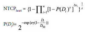
to a single dose. The Relative Seriality (RS) model was
used for calculation of the NTCP for late cardiac mortality.
The LKB NTCP and RS models used in this study
are briefly described below.[
Where TD50 is the 50% tolerance dose at 5 years;
EUD is the EUD; n and m are tissue-specific parameters;
Di is the dose element, and Vi is the volume element.
The parameters for complication of radiationinduced
pneumonitis are TD50=24.5, n=0.87.
RS model for late cardiac mortality:
Where D_50=TD50 (=52.3Gy); s is the RS factor
(=1), and ? is the maximum relative slope (=1.28).
The CLD is defined as the perpendicular distance
from the posterior edge of the tangential field to the
posterior part of the anterior chest wall in the middle
of the field. Maximum Heart Distance (MHD) is the
maximum distance of the heart contour to the posterior
field border measured in the beam's eye view of the
medial tangential fields.
Before calculation, DVH data were converted to the
dose biologically effective to 2 Gy per fraction irradiation,
using linear-quadratic model (α/β was equal to 3
Gy for both lung and heart). For the left lung, clinical pneumonitis of grade greater than 2 was used as endpoint
in NTCP calculation. In the case of heart, NTCP
calculation endpoint was excessive cardiac mortality in
the irradiated patients with the left-sided breast cancer.
The RS model coefficients used for lung and heart NTCP
calculation in this study were obtained from data analysis
of patients with breast cancer by Gagliardi et al.[
Statistical Analysis
Using the dose calculation data, we derived the DVH
curves of each OAR. For each patient and structure,
a cumulative DVH was exported from Eclipse with
a bin size of at most 5 cGy and imported into third
party program R studio. Here, NTCP calculations explained
below were performed using DVH metrics
vignette.[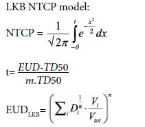
Data were compiled using Microsoft Excel and analyzed
using the Statistical Package for the Social Sciences
(SPSS) software version 20.0 (SPSS Inc, Chicago,
IL, USA). The mean and standard deviation of quantitative
variables was calculated. The mean between two
groups was compared using independent t-test. Quantitative
discreet variables were compared using the Chisquare
test. The correlation coefficient was estimated to
assess the relationship between CLD and MHD with
NTCP. P<0.05 was considered statistically significant
and p<0.01 was considered highly significant.
The present study attempted to evaluate dose-volume
histogram of heart, lung, and spinal cord in 3DCRT,
to analyze adherence of plan with QUANTEC
dose constraints. Mean dose for lung was 17.49 Gy, and
V20Gy was 39.88%. Mean dose for heart was 12.31 Gy
and spinal cord maximum dose was 0.78 Gy. However,
the test of significance observed no statistical difference
in the mean dose for heart, lung, and spinal cord
among patient receiving two-field or three-field radiation
therapy (p>0.05) (Table
In the present study, the average lung NTCP for radiation
pneumonitis was 3.11%. The risk of radiation
pneumonitis was observed to be significantly greater
in three-field radiation technique as compared to two
fields for lung (p<0.05). Hence, late cardiac mortality
probability for heart did not vary significantly between
two-field and three-field technique, but average heart
NTCP was 2.42%, as shown in Table
The NTCP in the ipsilateral lung revealed a significant
difference as the CLD increased from 3 cm to
4 cm (p=0.0012), as shown in Table
The present study observed the statistically significant
correlation of MHD and NTCP, that is, as the
MHD increased, the NTCP also increased in both twofield
and three-field radiation therapy (p<0.00001 and
p=0.000402), as depicted in Figure
MHD: Maximum heart distance; NTCP: Normal tissue complication probability.
CLD: Central lung distance; NTCP: Normal tissue complication probability.
In the present study, correlation of CLD and MHD
with dose-volume histogram was observed to be statistically
significant (p=0.001). For the breast the slope of
the V10Gy, V20Gy and V30Gy were nearly constant, as illustrated
in Figure
CLD: Central lung distance.
The analysis for the heart DVH was reflected
through the heart volume V25Gy plotted versus MHD.
The MHD was observed to be significantly correlated
with dose-volume histogram (p<0.001), as depicted
in Figure
MHD: Maximum heart distance.
RT has become the essential modality of treatment
in post-operative cases of locally advanced breast cancer.[
QUANTEC suggested <20% chance of symptomatic
pneumonitis with if <30% of lung received
20Gy (V20<=30%), whereas in the present study, V20Gy
was 39.88%. The mean dose in the present study was
17.49Gy. QUANTEC suggested 10% and 20% symptomatic
pneumonitis at the mean dose of 13 Gy and 20
Gy, respectively.
Similarly, for heart mean dose given in the present
study was 12.31 Gy, however, QUANTEC proposed 26
Gy, so it corresponds to QUANTEC constraint. Spinal
cord maximum dose is 0.78 Gy which is way lower than
QUANTEC constraint of 50 Gy for 0.2% myelopathy
rate. However, test of significance observed no statistical
difference in mean dose for heart, lung, and spinal
cord among patient receiving two-field or three-field
radiation therapy (p>0.05).[
In the present study, the average lung NTCP for radiation
pneumonitis was 3.11%. The risk of radiation
pneumonitis was observed to be significantly greater
in three-field radiation technique as compared to two
fields for lung (p<0.05). The rationale behind this effect
is due to the presence of the anterior supraclavicular
field, which elevates radiation dose in lung parenchyma
that lies beneath the chest wall. The findings of our
study are similar to the findings of study conducted by Noh et al.[
In the present study, late cardiac mortality probability
for heart did not vary significantly between two-field and
three-field technique, but average heart NTCP was 2.42%.
These findings were similar to the findings of study conducted
by Noh et al.[
Hurkmans et al.[
The CLD is commonly used as a simple parameter
to assess irradiated lung volume. We observed that as
the CLD increased from 3 cm to 4 cm, risk of organ
damage ipsilateral lung as revealed by NTCP increased
(p<0.01). Furthermore, a statistically significant correlation
of CLD and NTCP was established, as the
CLD increased, the NTCP also increased (p<0.001)
in three-field radiation therapy (p<0.001) but not in
two-field technique. This finding is suggestive of the
fact that chances of occurrence of pneumonitis in the
three-field radiation technique are highly dependent
on the CLD; probably, the supraclavicular field in the
three-field method increases the irradiated lung volume.
Noh et al.[
The MHD is a radiographic parameter that helps to
measure the irradiated heart volume for each patient.
Hurkmans et al.,[
In the present study, correlation of CLD with DVH
was observed to be statistically significant. For the
breast, the slope of the V10Gy, V20Gy, and V30Gy was nearly
constant. Das et al.[
In the present study, correlation of MHD with DVH
was observed to be statistically significant. The analysis
for the heart DVH was reflected through the heart volume V25Gy plotted versus MHD. The MHD was observed
to be significantly correlated with DVH. Das
et al.[
An accurate absolute NTCP model of excessive
late cardiac mortality and RP requires accurate clinical
data. However, several factors limit the accuracy
of these models. These include a long latency period
before clinical complications arise, the relatively low
incidence of radiation-induced cardiac complications
with newer RT techniques, and the relatively
high prevalence of ischemic heart disease in the general
population and the absence of good dosimetric
and volumetric data in retrospective studies. Although
the absolute NTCP values should be viewed
with caution, they must be fitted to clinical outcome
data and then are useful approximations of risk. This
study is limited in that it does not consider many
other risk factors of cardiovascular diseases, such as
a history of hypertension, smoking, and use of combined
cardiotoxic chemotherapy.
Peer-review: Externally peer-reviewed.
Conflict of Interest: All authors declared no conflict of interest.
Ethics Committee Approval: The study was approved by the Gandhi Medical College, Bhopal Ethics Committee (no: 3827-29/MC/IEC/2018, date: 30/01/2018).
Financial Support: None declared.
Authorship contributions: Concept - O.P.S., V.Y.; Design - O.P.S., V.Y., R.S.; Supervision - S.Y., P.K.P.; Funding - V.Y., S.Y.; Materials - R.S., H.U.G., V.T.; Data collection and/or processing - R.S., M.C., S.S.; Data analysis and/or interpretation - R.S., V.Y., P.K.P.; Literature search - O.P.S., V.Y., R.S.; Writing - R.S., V.Y.; Critical review - O.P.S., V.Y.

