

METHODS
To simulate rando phantom geometrically, arms and legs created by rice are added to rando phantom
and were immobilized by a vacuum bed. The computer tomography images of the lower and upper
region of the phantom with a slice thickness of 3 mm were acquired. Thermoluminescent dosimeters
(TLDs) were placed entrance-exit, midpoint doses of lens, lungs, and kidneys onto a rando phantom.
The treatment plans were prepared and irradiated with helical tomotherapy technique. Results of the
measurements and treatment planning system (TPS) doses were compared. All procedures were repeated
3 times and averaged. TPS and TLD doses were evaluated by Wilcoxon test.
RESULTS
In the plans prepared with the TPS, a homogeneous dose distribution was obtained with a homogeneity
index of 0.16 for the upper body and 0.04 for the lower body. When the calculated critical organ doses
were compared with the measured organ doses, there was no statistically significant difference between
them.
CONCLUSION
It was seen that saving critical organs and achieving desired dose distribution are possible without blocks
in tomotherapy. Doses calculated from TPS and measured by TLD matched.
Keywords: Helical tomotherapy; rando phantom; thermoluminescent dosimeters; total body irradiation
In TBI s, it becomes hard getting homogenous dose distribution due to inhomogeneous body shape and tissue density variations. The main goal of the radiotherapy is saving critical organs while achieving a homogeneous dose distribution in the target volume.
There has been a wide range of TBI treatment techniques
from the past to now. While classical TBI treatments
were performed with Co-60 and long source skin
distance (SSD) Linac in the past, today treatments are
applied using advanced technologies such as volumetric
modulated arc therapy (VMAT) and tomotherapy.
When old technologies are preferred, external shielding
blocks or compensators are used and for long SSD
Linac treatments, big treatment room size is needed in
the clinic. Treatment room size, patient comfort, and
treatment times are important factors to feel the need
for developing technologies for TBI treatments. The big
treatment room size, time taking factors like shielding
blocks, and compensators are not needed in the new
technologies. Treatment time is also another important
advantage of tomotherapy and VMAT. Recently,
the most preferred and effective two technologies used
in TBI treatments which are as follows: Tomotherapy
and VMAT. Tomotherapy is a new and one of the most
effective technologies used for TBIs. It is a 6 MV machine
that is a combination of computed tomography
(CT) and intensity modulated radiation therapy. Since
it gives MVCT images before each treatment, appropriate
patient localization, and beam delivery can be
achieved in tomotherapy.[
In TBI treatments, the most common dose fraction
schedule is12 to 15 Gy given in 8 to 12 fractions over 4
days, with 2 to 3 treatments daily.[
In this study, it was aimed to investigate TBI planned
target volume (PTV) coverage and organ doses using
helical tomotherapy. PTV dose and critical organ doses
such as lens, lungs, and kidneys were measured by TLD
on the phantom. PTV and critical organ doses on the
treatment planning system (TPS) and measured TLD
doses were compared.
The arms and legs created using rice were added
to the male phantom (CIRS, Computerized Imaging
Reference Systems Inc. Virginia, USA) to simulate the
whole body geometrically. The male phantom with
arms and legs was immobilized by a vacuum bed. The
CT images of the lower and upper region of the phantom
with a slice thickness of 3 mm were acquired using
Philips Big Bore CT (Philips Medical Systems, Cleveland,
OH, USA). The CT datasets were transferred to
the TPS. Figure
CIRS: Computerized Imaging Reference Systems; CT: Computed
tomography.
In Volo TPS (Accuray, Incorporated, Sunnyvale, CA), PTV was created as the entire body with 5 mm inner margin from the skin, and then, lungs, lens, and kidneys were contoured with a margin of 3 mm from the PTV. Dose constraints for organs at TPS were; mean coverage of 90% PTV is 12 Gy, maximum lens doses are 5 Gy, mean lung doses are 8 Gy, and mean kidney doses are 7 Gy. As planning parameters, 5.054 cm field width, 2.0 modulation factor, 0.415 pitch value, dynamic jaw mode, and helical beam mode were prescribed to TPS. The upper and lower part of the body was planned separately and united in MIM software (MIM Software, Cleveland, OH) to control hot and cold spots in intersecting areas.
45 number of GR-200A (PTW, Physikalisch Technische Werkstätten, Freiburg, Germany) with a diameter of 4 mm and 0.8 mm-thickness disc TLD were used. In this study, three groups of TLD were used. The standard deviations for each group of TLD were <2%. Each group of TLDs was calibrated to 1 Gy in a solid water equivalent phantom with a 10×10 cm2 field size at a depth of 5 cm and a source-axis distance is 100 cm. In the first part of the study, TLDs were placed into the male phantom and irradiated using TomoTherapy-HDA Treatment System (Accuray, Incorporated, Sunnyvale, CA) with helical tomotherapy technique. TLDs were read out with Fimel LTM Reader. Entrance-exit, midpoint doses of lens, lungs, and kidneys were measured. This procedure was repeated 2 times and average values were considered. The second part of the study, it was aimed to make a comparison of planned and measured doses. The TPS and TLD doses were evaluated by Wilcoxon test.
Organ doses for upper body plan at VoLo TPS are; PTVmax=14.45 Gy, right lungaverage=7.61 Gy, left lungaverage =7.70 Gy, right kidneyaverage=6.70 Gy, left kidneyaverage =6.51 Gy, right lensmax=2.94 Gy, left lensmax=3.18 Gy, and externalmax=14.45 Gy.
For the lower part of the body, Figure
Lower part doses are; PTVmax=13.12 Gy and external max=13.12 Gy.
DVH: Dose volume histogram.
After evaluation of upper and lower body plans,
two plans were brought together in MIM software program to check hot or cold spots at intersecting areas.
Figure
DVH: Dose volume histogram.
All planning values were in desirable range for TBI treatments. To ensure this, dose homogeneity index (HI) was calculated with the shown formula. According to ICRU 83 HI close to zero means, PTV dose distribution is homogeneous.[12]
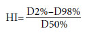
Calculated HI values in this study were for the upper body; 0.16, and for the lower body; 0.04. Results show that in this study, TPS provides homogenous dose distribution for PTV in TBI treatments.
In TBI treatments, since the target body is very large, time is another important factor for both patients and treatment team. To decrease, treatment time makes the process more comfortable and repeatable. One more advantage of tomotherapy is that it is very effective in decreasing treatment time in TBI treatments. In this study, treatment time for the upper body was 12.9 min, and for the lower body was 13.5 min. These numbers are very ideal for TBIs.
TPS Reading Doses and TLD Measured Doses
Comparison
After getting TPS values, 15 numbers of TLDs were
placed onto the male phantom to check TPS doses and
measured doses. TLDs were placed on the lens, head
midpoint, thyroid entrance-exit points, lungs midpoints,
between two lungs, kidneys midpoints, kidneys
entrance-exist points, two plan intersecting areas, and
between two feet. TLDs were placed at these points and irradiated 2 times. Average of two irradiation values
was calculated and made a comparison between
planned and measured doses of organs for one fraction.
The comparison was made with Wilcoxon test
and as shown in Table
In TBIs, it becomes hard getting homogenous dose distribution due to inhomogeneous body shape and tissue density variations. The main goal of radiotherapy is to save critical organs while achieving a homogeneous dose distribution at the target volume. When it comes to deciding for TBI treatment method, some other crucial points must be considered. These considerations are treatment time, planning time, repeatability, and comfortableness for both patient and team.[2] When all these considerations are taken into account, tomotherapy stands out as the most ideal treatment technic for TBIs. The aim of this study is to investigate the suitability of tomotherapy for TBI and the coherence of planned and given doses.
For TBI treatments, 80% of the given dose are acceptable
as the lungs" tolerance dose. 7-10 Gy lung
mean dose is admissible and the midpoint of the
lungs is seen as the reference point. Lens dose is admissible
under 5 Gy for all treatments. These are dose
limits for TBI in practice and measured doses in this
study are in the desired range of limits. Tomotherapy
is very effective for saving critical organs while
achieving homogeneous dose distribution in PTV. The skin dose in TBI should not be under 90% of the
given dose[
Several publications demonstrated the practicality of
tomotherapy for TBI. Penagaricano et al. made a study
with four AML patients to investigate clinical feasibility
of helical tomotherapy for TBI. TBI prescription was set
that 80% of the clinical target volume received 12 Gy in
six fractions, at two fractions per day. 3 mm CT images
were used for plan and lungs and kidneys were saved as
critical organs. 5 cm jaw width, 2.0 pitch value, and 0.287
modulation factor were used for the plan parameters.
Planning average doses of OAR were set not to exceed
8 Gy and 10 Gy to the lungs and kidneys, respectively.
In the treatment process planned and delivered
CTV doses ranged from 12.1 to 12.4 Gy and 11.9-
12.3 Gy, average planned and delivered lung doses
ranged from 6.6 to 7.4 Gy and 6.5 to 7.4 Gy, respectively.
Average planned and delivered left kidney
doses ranged from 7.4 to 8.7 Gy and 7.2 to 8.6 Gy, respectively.
Average planned and delivered right kidney
doses ranged from 7.5 to 8.6Gy and 7.3 to 8.5 Gy. The planned and delivered dose HI ranged from 0.91
to 0.94 and from 0.90 to 0.95. All results convince
feasibility of tomotherapy for TBI.[
Gruen et al.[
Hui et al.[
In another study conducted by Zhuang et al.,[
Sarradin et al. conducted a study with 11 patients
who were treated between August 2014 and January
2016. The total dose was 12 Gy in six fractions in 3 days.
The median age was 31 years, range from 18 to 57 years.
The median D98% of PTV was 11.5 Gy, ranging from
6.6 to 11.9 Gy. The average of the mean dose to the lungs
was 8.7 Gy, ranging from 8.5 to 9.3 Gy. The mean dose
for the junction area was 12 Gy, ranging from 11.9 to
12.1 Gy. In the study, they saw that no patient had radiation
pneumonitis. According to this study, Sarradin
et al. affirmed the efficiency of helical tomotherapy in
TBI treatments in terms of sparing organs at risk while
achieving uniform dose coverage at target volume.[
Sun et al.[
Inhomogeneous body shape and tissue density
variations make it hard to achieve uniform dose distributions
in TBI treatments. The most crucial consideration
of radiotherapy is saving critical organs meanwhile
achieving homogeneous dose distribution at the
target volume. Demonstrated studies show that tomotherapy
is a very effective method in TBI treatments
since it achieves the main goal of radiotherapy and it is
comfortable for both patient and user.
Peer-review: Externally peer-reviewed.
Conflict of Interest: All authors declared no conflict of interest.
Ethics Committee Approval: The study was approved by the Istanbul University, Institute of Oncology Ethics Committee (no: 338172, date: 22/09/2016).
Financial Support: None declared.
Authorship contributions: Concept - H.B.B., G.U.A.; Design - H.B.B., G.U.A., İ.K.Ç.; Supervision - H.B.B., G.U.A., C.K.A.; Funding - H.B.B., G.U.A., İ.K.Ç.; Materials - H.B.B., G.U.A.; Data collection and/or processing - H.B.B., G.U.A., İ.K.Ç.; Data analysis and/or interpretation - H.B.B., G.U.A., C.K.A.; Literature search - G.U.A., C.K.A.; Writing - H.B.B., G.U.A., İ.K.Ç.; Critical review - H.B.B., G.U.A., C.K.A., İ.K.Ç.

