

METHODS
A modular structure of the metallic vaginal speculum, which we use in our routine practice, can be easily
separated from the handle of the speculum, has been designed and produced with a 3D printer from
PEEK material with a layer thickness of 200 µ. Dosimetric measurements of speculum tongue made of
PEEK material were made using two different dosimetric methods (three different techniques as the
treatment planning system ACUROS BV and TG43 algorithms, and the Radiochromic Film Dosimetry
system [Gafchromic: EBT3]).
RESULTS
The values obtained by all three methods were correlated with each other and the speculum did not affect
negatively dose distribution in the dose-distance curves. The dose values obtained by using ACUROS
BV and TG43 algorithms at the Dwell position and the dose values obtained in the Gafchromic dosimetry
measurements made at these points were found to be correlated with each other (ACUROS BV vs.
TG43 p<0.001, ACUROS BV vs. EBT3 p=0.008, TG43 vs. EBT3 p=0.006).
CONCLUSION
When all the measurement results were examined, it was seen that the values obtained were correlated
with each other, and no negative effect of the speculum on the dose distribution was observed in the
dose distance curves.
Keywords: 3D printer; dosimetry; polyetheretherketone; vaginal speculum
The purpose of brachytherapy treatment is to place
radioactive sources near or inside the tumor. This ensures
not only increased dosage in the targeted tissues
with tumors and improvement of overall survival
through local control but also reduced dosages for
the bladder, rectum, sigmoid, and other organs at risk
(OARs) beyond or near the target.[
Brachytherapy procedure generally lasts 1.5-2 h
and might be implemented under general or spinal anesthesia
as the process of placing the applicators inside
the cervix, endometrium or tumor may be accompanied
by pain or anxiety. During this process, the patient
is supposed to lie down and hold still. In brachytherapy
applications, it is crucial to select the correct applicator,
place it correctly, and then, keep OARs away from
the radioactive source. For this purpose, a stainlesssteel
vaginal speculum that is available in the market
and routinely used is fitted before the applicators are
placed into the cervix and uterus. Then, the packing
of the vagina is performed using the gauze dipped into
normal saline to keep away the bladder in the front and
the rectum at the back. After this process is completed,
the vaginal speculum which has a modular structure
is split into two parts the upper and lower parts, and
pulled outside of the vagina.[
Polyetheretherketone (PEEK) is one of the outstanding
polymers in the semi-crystalline thermoplastic polymer
class. Today, PEEK is available in various forms such
as fiber, film, resin, fabric, cable, membrane, and composite.
Chemically, the PEEK polymer consists of repeating
units of one ketone group and two either groups,
and it is durable and biologically compatible thanks to
its fully aromatic, highly stable, and linear structure that
contains only carbon, hydrogen, and oxygen atoms.[
The starting point of this study is the possibility of
replacing the metal speculum with PEEK so that it can
be left in place during gynecologic brachytherapy procedures.
Hence, this project aims to modify the vaginal
speculum that is currently available in the market
and routinely used in daily practice by producing the
lower and upper blades of the vaginal speculum using
a PEEK material and performing dosimetric measurements,
and testing the feasibility of the resulting modification
for brachytherapy applications.
PEEK: Polyetheretherketone.
Design and Production of the Test Apparatus
For dosimetric measurements regarding the radiation
permeability of the produced speculum blades, a phantom
apparatus was designed and produced with a width
of 8×10 cm and a height of 12 cm using the glass material.
A guide and locking mechanism were produced from
polylactic acid using an Intamsys Funmat HT 3D printer
to ensure that the speculum blades and radioactive source
can be secured inside the apparatus (Fig.
Performing Dosimetric Measurements
Two different dosimetry methods (treatment planning
system (TPS) and Radiochromic Film Dosimetry
System (GAFChromic: EBT3)) were used to perform
dosimetric measurements of the blades of the vaginal
speculum produced from the PEEK material, and the
values calculated using these two methods were compared
to each other.
Measurements Made using the TPS
Radiochromic Film (GAFChromic Film) Dosimetry
Calibration and Measurements Made
The values obtained were used to draw dose versus
OD calibration curves. The calibration curves were
later used to measure dose values from the measured
OD values of the tarnishing on the film as a result of
experimental irradiation.
Following the calibration of radiochromic films, the
test apparatus, described above, was used to perform the
irradiation of radiochromic films on the brachytherapy
device at the sides with and without the speculum.
The speculum blades manufactured from the PEEK
material were placed inside the measurement apparatus
and their CT images were taken for planning purposes.
The CT sections obtained were transferred, and
two different brachytherapy applications were prepared
using ACUROS BV and TG43 algorithms at the same
source position and for the same duration (Figs.
In the brachytherapy planning made using two different
algorithms, the irradiation was performed, and
film dosimetry was conducted without changing the test
apparatus and ensuring that GAFChromic films were
placed to align them with the source. The dose values
obtained from the TPSs using two different algorithms
and the two different irradiations for the duration obtained
from the planning and the mean values of two
different assessments for each are given in Figure 7.
Statistical Analysis
The Eclipse TPS (Version: 15.6, Varian Medical Systems,
Palo Alto, United States of America) was used
as the TPS. In dosimetric comparisons, computed
tomography (CT) (Siemens Somatom AS, Siemens
Healthineers, Erlangen, Germany) of the test apparatus was taken first. The guiding tube (bar) with code
no. GM11011180 of the cylinder applicator with code
no. GM11011100 of the Varian GammaMed Plus iX
brachytherapy device (Varian Medical Systems, Palo
Alto, United States of America) was placed at the center
of the testing apparatus. To enable measurements with
and without the speculum in the test apparatus, one
blade of the speculum was placed 2.3 cm away from
the applicator to the left of the source and no speculum
blade was placed to the other side. This point was chosen
because it is the distance at which a dose of 3 Gy is
delivered in the TPS. Here, the aim was to perform dosimetric
measurements from different directions and
compare them. After these processes, water was placed
inside the test apparatus as representative of soft tissue.
For dose calculations, the Acuros BV calculation
algorithm (Varian Medical Systems, Palo Alto, United
States of America), which takes Hounsfield units in
TG43 and CT images, into consideration, was used.
Dose values at the specified points at the sides with and
without the speculum (the points where GAFChromic
films will be placed) on the images sets obtained from
CT were recorded using the beam data and calculation
algorithm belonging to 192lr (t1/2: 73.827 days) previously
loaded into the system. On both sides, the points
were set 5.02 (LEFT), 4.07 (RIGHT), 4.07 (DOWN),
and 3.95 (UP) cm away from the source and 5 cm away
from the table plane to minimize the scattered photon
effect on the measurement results.[
In the dosimetric measurements made using Radiochromic
film (Ashland Specialty Ingredients, Bridgewater,
NJ, United States of America), first, the calibration
curves of these films were obtained. To this end,
the Varian GammaMed Plus iX model brachytherapy
device (Varian Medical Systems, Palo Alto, United
States of America), located at the Radiation Oncology
Department of the Faculty of Medicine Hospital of
Eskişehir Osmangazi University and contains a 192Ir
radioactive source, was used. For the calibration of the
radiochromic films, the films were cut into a size of
2.5 cm×2.5 cm. The films were cut using scissors and
the films were handled using gloves to avoid any stains
on film surfaces. The cut radiochromic films were categorized into two groups as irradiation and control.
The irradiation group consisted of the films to be irradiated
at the brachytherapy device. The films in the
control group were not irradiated, and they were used
to eliminate the tarnishing resulting from the ambient
radiation in radiochromic films during the time between
irradiation and scanning. Both groups of films
were read in an Epson Expression 11000XL scanner
(Epson Seiko Corporation, Nagano, Japan) to get a
minimum lateral scattering effect before moving to
the irradiation process.[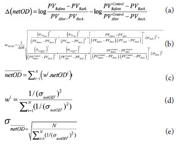
Categorical variables were described as counts and percentages
(%), whereas continuous variables were described
as means (±standard deviation). The correlations
(r values) were assessed using Pearson's correlation coefficient.
All tests of significance were two-tailed with p<0.01.
The correlation of the doses obtained using the
ACUROS BV and TG43 algorithms at the points determined
at the level of the Dwell position of the radioactive
source and obtained from these points using
the GAFChromic dosimetry method was performed,
and it was found that the values obtained using each of
three methods were correlated to each other (p<0.001
for ACUROS BV vs. TG43, p=0.008 for ACUROS
BV vs. GAFChromic Film, p=0.006 for TG43 vs.
GAFChromic Film) (Table
Two different dosimetry methods (TPS and Radiochromic
Film Dosimetry System [GAFChromic:
EBT3]) were used to perform dosimetry, and it was
found that the values calculated using the three methods
were correlated to each other. Examination of the
dose-distance curves revealed that the modified speculum
did not have any adverse contribution to the dose
scattering, and it has to be supported by trials to mention
that the speculum blades produced from the PEEK material can be used for brachytherapy applications.
However, there are different measurement methods in
terms of dosimetry, and in this study, film irradiation
was performed at two different times to measure the
dose obtained from two different planning algorithms,
and the measurement of each film was performed twice.
In a total of four assessments, different results were obtained.
For this reason, mean dosimetric measurements
were given in this study. In the literature, an uncertainty
of approximately 7% for lower doses and above 8% for
higher doses was reported, and lower rates of uncertainty
were reported using different dosimetry methods
such as Metal Oxide Semiconductor Field Effect Transistor
and Optically Stimulated Luminescence.[
Peer-review: Externally peer-reviewed.
Conflict of Interest: All authors declared no conflict of interest.
Financial Support: This study has been supported by the Eskişehir Osmangazi University Scientific Research Projects Unit with project number TSA-2021-2143.
Authorship contributions: Concept - A.Ö.; Design - A.Ö.; Supervision - A.Ö.; Data collection and/or processing - A.Ö., K.D.; Data analysis and/or interpretation - A.Ö., K.D.; Literature search - A.Ö., K.D.; Writing - A.Ö., K.D.; Critical review - A.Ö., K.D.

