Introduction
Cervical cancer is a prevalent malignancy in women,
ranking fourth worldwide based on the 2020 Global
Cancer Observatory statistics (GLOBOCAN). This disease
ranks 12th in our country, with an incidence rate
of 4.8 per thousand, as reported in the literature.[,]
Radiotherapy represents a pivotal treatment modality,
especially in cases with lymph node metastasis. As
chemotherapy technology evolves, radiotherapy has
emerged as the standard treatment approach. In cases
where metastasis has occurred in the paraaortic lymph
nodes, careful evaluation of all volumes and organs at
risk is critical to ensure treatment success with minimal
side effects while providing a uniform dose distribution
within the target volume.[-] Thankfully, advancements
in radiotherapy techniques have improved treatment
outcomes. While three-dimensional conformal
radiotherapy (3DCRT) was commonly used in the past,
the latest techniques such as intensity-modulated radiotherapy
(IMRT) and volumetric-modulated radiotherapy
(VMAT) have grown in popularity due to their ability
to offer fewer fractions, shorter treatment times, and
high fraction doses.[-] These techniques have also
shown promise in studies involving gynecologic cancers,
as they cause less damage to normal tissues, reduce
treatment times, and require fewer monitor units.[,]
Recent research has focused on comparing IMRT
and VMAT techniques for simultaneous integrated
boosts (SIB) in various anatomical sites. This is because
the clinical use of VMAT has increased.[,] However,
there is still a lack of comprehensive studies evaluating
these techniques, particularly in cases of cervical cancers
with paraaortic lymph node (PALN) metastases.[,]
The focus of our research was on conducting a comparative
analysis of the dosimetric aspects of IMRT and
VMAT plans under SIB conditions to determine the
optimal treatment approach for patients undergoing
radiotherapy. We sought to evaluate the effectiveness
of SIB-VMAT and SIB-yttrium-90 radioembolization
(SIB-YART) treatment planning methods for cervical
cancer patients, focusing on the Planned Target Volume
(PTV) and doses received by organs at risk. To achieve
this, we conducted a statistical analysis of the dose volume
histograms (DVH) generated by both techniques,
comparing their respective dosimetric properties.
Methods
This research involved 10 patients who had been diagnosed
with cervical cancer with PALN metastasis and had completed their treatment, with the Department"s
consent. Written consents required for the study were
obtained in compliance with the Declaration of Helsinki,
and approval was granted by the local ethics committee
(Decision No: 24-2.1T/75).
In order to ensure comprehensive treatment, all patients
underwent a pelvic-paraaortic field plan. A radiation
oncologist utilized computed tomography (CT)
scans of the pelvic region from ten patients to create
the PTV. The plan incorporated definitions of the gross
tumor volume (GTV), clinical target volume (CTV),
internal tumor volume (ITV), and contours relating to
critical organs (OAR), for instance, bladder, bowel, femoral
heads, kidneys, liver, rectum, sigmoid, and spinal
cord. The PTV was designed with a margin of 1.0 cm
for the CTV and 1?1.5 cm for the ITV, allowing organ
movement while limiting the dose to normal tissues.
The planned dose for the elective volume was 45
Gy delivered in 25 fractions of 1.8 Gy each, and the
integrated boost volume was planned to administer
55 Gy over the same 25 fractions of 2.2 Gy simultaneously.
The Eclipse Treatment Planning System was
utilized to create SIB-IMRT and SIB-VMAT plans
with 6-MV photon beams from a Varian TrueBeam
STX linear accelerator in California, USA. The IMRT
plans were optimized using the dose volume optimizer,
while the VMAT plans utilized the progressive
resolution optimizer. Both plans were calculated using
the AcurosXB planning algorithm (version 1610).
In the creation of IMRT plans, the sliding window
algorithm application method was employed, utilizing
nine planar equal fields placed at varying degrees
around the patient. For the VMAT plan, two coplanar
arcs of 360 degrees were utilized with the same isocenter.
Both plans were normalized to obtain a minimum
of 95% of the prescribed dose and thoroughly
evaluated based on the resulting parameters. Both
plans utilized identical planning targets for every patient.
The numerical target table was populated with
the same objectives for optimizing IMRT and VMAT
plans. The primary focus during dose delivery was to
guarantee that 95% of the prescribed dose reached the
PTV, followed by minimizing the dosage to critical organs
like the kidneys, and finally, administering lower
doses to other OAR structures to reduce the dosage to
critical organs to the fullest extent possible. The plans
for both techniques were developed to yield optimal
outcomes for 10 patients. DVH was used to analyze
dose values to evaluate the quality of each plan for
PTV (elective and SIB). These values included the
minimum, maximum, and mean doses for OAR, and additionally assessed the volume that receives 95% of
the prescribed dose and the maximum doses received
by 2%, 50%, and 98% of the volume. From these values,
the literature determined the homogeneity index
(HI) and conformity index (CI) for individual plans.
A small HI value indicates a consistent dose distribution
across the volume, while a CI value nearing 1
indicates a high degree of conformity in the delivery
of the dose to healthy tissues surrounding the target
volume. The formulas for these expressions are:
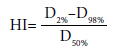
Regarding radiation dosage, D2% is the dosage in
the target volume, also known as the 2% target volume
dose. D98% refers to the minimum dosage within
the target volume, or the 98% target volume dose.
Meanwhile, D50% pertains to the average radiation
dose received in a tumor, which encompasses up to
fifty percent of the volume designated as the target. If
the HI value is low, this indicates that the dose distribution
within the volume is homogeneous.
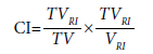
The TVRI refers to the volume intended to receive
radiation treatment and is surrounded by a reference
isodose. Meanwhile, TV pertains to the target volume,
while the VRI parameter defines the volume of the reference
isodose. A CI value nearing 1 indicates a reduced
radiation dose to the tissues surrounding the target
volume, as supported by previous studies.[,,]
In order to compare the OAR sparing ability
of SIB-IMRT and SIB-VMAT plans, we analyzed
the volumes of the bladder, bowel, femoral
heads, liver, rectum, sigmoid, and kidneys using using
V70(%), V195(cc), V15(%), V30(%), V40(%), V40(%),
V55(%),V32(%) V30(%), V20(%). Additionally, we examined
mean dose values of 18 Gy and 32 Gy for kidneys
and liver, and the maximum dose limit of 45 Gy
for the spinal cord. Previous research has indicated
that VMAT may produce lower MU values compared
to IMRT due to less radiation leakage through collimator
leaves and reduced neutron production at high
energies. As such, we recorded technique-specific
MU values for our study aims to assess the efficacy
of each technique.[,,] We utilized the Wilcoxon
paired two-sign rank test to perform a statistical
evaluation on nonparametrically distributed data.
A significant difference was indicated if the p-value
equaled or fell below 0.05 (p≤0.05).[]
Results
All 9 patients achieved the SIB targets for PTV coverage
and OAR sparing with the SIB-IMRT and
SIB-VMAT plans. However, the IMRT plan for one
patient failed to achieve its target due to inadequate
use of the 9 fields in treatment. On the other hand,
the VMAT plan successfully achieved the SIB targets.
Each plan was designed to be clinically feasible. The
techniques showed statistically significant differences
in MU, kidney, and liver values.
The comparison results of Tables 1 and 2 depict
SIB planning utilizing IMRT and VMAT techniques.
Table 1 focuses on the parameters of SIB-IMRT and
SIB-VMAT for PTV55 coverage, while Table 2 presents
the performance of the two techniques regarding OAR
preservation. In Table 1, SIB-VMAT outperforms SIBIMRT
in terms of MU (545.91), CI (0.74), and HI
(0.04), although the HI results are not statistically significant.
Conversely, Table 2 indicates that SIB-VMAT
is significantly superior to SIB-IMRT in preserving
OAR in the right kidney V30(%), V20(%), left kidney
V30(%), V20(%), and liver Dort(Gy) values with 9.83%,
9.26%, 8.15%, 7.64%, and 7.74Gy values, respectively
Table 1 The mean data acquired for the comparison of
SIB-IMRT and SIB-VMAT techniques in 10 patients
for PTV55 dose, along with p-values from the
Wilcoxon-signed rank test
Table 2 Mean dose for organs at risk (OAR) between SIB-IMRT and SIB-VMAT in 10 patients
along with p-values from the Wilcoxon-signed rank test
In Figure 1, a and b are the isodose distributions of
both the SIB-IMRT and SIB-VMAT plans for a patient
in the transversal section (top), along with the isodose
distributions of the same patient's treatment plans in the
frontal section (bottom). Additionally, Figure 2 showcases
the PTV and OAR DVRs of the pertinent parameters
in the patient"s SIB-IMRT and SIB-VMAT plans,
highlighting the defined criteria in varying colors.
Fig. 1. The Comparison of isodose distribution in a specific patients SIB-IMRT (a) and SIB-VMAT (b). Blue and green-segmented volumes receive 45 and 55 Gy,
individually. Dark blue and light green lines represent 45 and 55 Gy, individually. Transverse (top) and frontal (bottom) sections, respectively: Y: -11, and Z:
-2.53 cm shown in the head-first-supine position.
SIB-IMRT: Simultaneous Integrated Boost-Intensity Modulated Radiation Therapy; SIB-VMAT: Simultaneous Integrated Boost-Volumetric Modulated Arc Therapy.
Fig. 2. The Comparison of Dose-volume histogram (DVH) depicting the associated organs and target volumes in the
SIB-IMRT (triangles) and SIB-VMAT (squares) plan, corresponding to the selected patient case shown in Figure 1,
presenting in various color category for the parameters of each PTV and OAR.
SIB-IMRT: Simultaneous Integrated Boost-Intensity Modulated Radiation Therapy; SIB-VMAT: Simultaneous Integrated Boost-Volumetric
Modulated Arc Therapy; PTV: Planned Target Volume; OAR: Contours relating to critical organs.
Discussion
Cervical cancers are highly treatable, and thanks to advances
in technology and medicine, survival rates are
on the rise. Radiotherapy is a critical element in treating
cervical cancer, and treatment plans are established according
to the stage of the disease and the extent of lymphatic
spread.[] This study aimed to compare treatment
plans using the SIB technique with those using
IMRT and VMAT techniques in 10 patients with cervical
cancer and PALN metastasis. Precise planning and
dosimetric measurements are crucial in radiotherapy
treatments, and techniques like IMRT and VMAT take
into account the structure of the target area to minimize
side effects while regulating radiation doses. Studies
have shown that SIB-assisted VMAT and IMRT treatment
plans produce more favorable outcomes.[,]
The preferred parameters to evaluate the two techniques
are HI, CI, and MU.[,,] Wu et al.[] conducted
a comparison of 2-arc VMAT and 9-field IMRT
techniques for 20 patients, using dose values of 45 and
50.4 Gy. They observed a notable disparity between the
techniques in HI value (p=0.036). On the other hand,
Vergalasova et al.[] did not identify a statistically
significant disparity in HI criteria in their study comparing two techniques for 20 patients (p=0.55). In our
research, we observed that the SIB-IMRT technique
showed superiority with an HI value of 0.04, although
no significant discrepancy was found for HI (p=0.08).
In Wu et al.[]'s study comparing the same techniques,
they obtained a CI value close to our IMRT result
(0.77) with the prescription dose values they chose (45
Gy and 50.4 Gy) and established a significant difference
among the techniques (p=0.016). In a study carried out
by Cheng et al.[] with 10 patients, the CI criterion revealed
a significant difference (p=0.001). Upon analyzing
the CI values, researchers found that the SIB-IMRT
technique was inferior with an assessment of 0.74,
and there was a significant variance compared to the
results of the SIB-VMAT technique (p=0.02). Wu et
al.[] discovered that the VMAT technique exhibited
superiority in the MU value, and there was a statistically
significant difference observed when compared to
other techniques (p=0.001). Meanwhile, Vergalasova et
al.[]'s study noted a statistically significant difference
for the MU criteria (p=0.00009) when comparing the
two techniques. In our research, we observed a significant
difference when analyzing the MU values between
the SIB-VMAT and SIB-IMRT techniques, with the
former proving statistically superior (p=0.005).
In a comparative study conducted by Riou et al.[]
it was found that the SIB-VMAT technique was superior
for bladder dose limit values in 10 patients. Conversely,
no statistically significant difference was found between
the SIB-VMAT and SIB-IMRT techniques. Similar findings
were reported by Vergalasova et al.[] who found
no statistically significant difference in bladder criteria
in their study (p=0.19). When we look at the bladder
values obtained in our assessment, we identified no significant
difference between SIB-VMAT and SIB-IMRT
(p=0.13). In the QUANTEC protocols examined, the
bowel limit was determined to be <195 cc.[] The exact
limits were applied in this study, and no significant difference
was observed in the comparison of bowel doses
(p=0.13). The RTOG protocols reviewed, and the dose
limits for the right-left femoral head were determined
as <15% and <15%, respectively.[] In this study, the
dose limits were determined in the same way, and in the
comparison made for SIB-IMRT and SIB-VMAT, the
values for the right femoral head were 10.79 and 10.94,
respectively, with no significant difference for p=0.16,
and the values for the left femoral head were 9.76 and
11.62, respectively, with no statistically significant difference
(p=0.24). Wu et al.[] found that the VMAT
technique showed superiority for the mean left-right kidney values with a significant difference between the
techniques (p=0.001, p=0.001, respectively). However,
Vergalasova et al.[] identified no significant discrepancy
in mean left-right kidney values. In the comparison
of mean left-right kidney doses between SIB-VMAT
and SIB-IMRT, no notable difference was found for the
left kidney, while a significant difference was found for
the right kidney doses (p=0.17, p=0.39, respectively).
In the examination of mean left-right kidney doses
between SIB-VMAT and SIB-IMRT, no significant difference
was established for the left kidney, while a significant
difference was found for the right kidney doses
(p=0.07, p=0.05, respectively). In the examined QUANTEC
protocols, the liver limit value was determined as
<32 Gy.[] In this study, the same limits were used, and
there was no statistically significant difference between
the two techniques for liver mean doses (p=0.09). Wu et
al.[] found that the VMAT technique was superior for
rectum value, and there was a significant difference between
the techniques (p=0.001). Cheng et al.[] found
a significant difference between the rectum values of
the two techniques in their study (p=0.001). In our
research, although there was no significant difference
between the two techniques for rectal values, percent
doses showed superiority in favor of the SIB-VMAT technique (p=0.95). In their comparison study of 8
patients, Jin et al.[] found the SIB-VMAT technique
superior for spinal cord dose limit values, and a significant
difference was found between the two techniques
(p=0.04). However, in our study, dose limits were determined
similarly, though no statistically significant
difference was detected in the comparison made for the
spinal cord (p=0.09). Based on the OAR dose data collected
in the study, the SIB-VMAT technique demonstrated
superiority in the average values of the right kidney,
left kidney, and liver, while remaining within the
specified limits. A significant difference was observed
between the two techniques. The SIB-IMRT technique
showed superiority in the average value of the left kidney,
and a significant difference was also detected.
When choosing between the SIB-IMRT and SIBVMAT
techniques, it is important to consider various
factors, such as the patient's condition and treatment
duration. Although VMAT has advantages in
terms of treatment duration, it requires more time
for treatment planning and quality assurance. However,
some studies suggest that quality assurance
plans are quicker with VMAT.[,]
Conclusion
Our study revealed that both VMAT and IMRT techniques
exhibit distinct advantages and disadvantages.
Therefore, the selection of the most suitable technique
should be based on the patient"s condition and treatment
planning. Our current research unveiled that although
there was a significant difference in SIB-IMRT and SIBVMAT
plans among 10 patients, both techniques provided
similar dosimetric results. Accordingly, it is crucial
to consider factors, particularly the patient's condition,
clinical equipment, and the expertise of doctors and
physicists, when selecting the appropriate technique.
Our results indicate that both SIB-IMRT and SIB-VMAT
techniques are effective in treating cervical cancer.
Ethics Committee Approval: The study was approved by
the Ege University Medical Research Ethics Committee (no:
24-2.1T/75, date: 22/02/2024).
Authorship contributions: Concept - İ.A.E., H.E., Z.Ö.;
Design - İ.A.E., H.E.; Supervision - İ.A.E., H.E., N.O.,
Z.Ö.; Funding - İ.A.E., H.E.; Materials - İ.A.E., H.E.; Data
collection and/or processing - İ.A.E., H.E.; Data analysis
and/or interpretation - İ.A.E., H.E., N.O.; Literature search
- İ.A.E., H.E., N.O.; Writing - İ.A.E., H.E., Z.Ö.; Critical
review - İ.A.E., H.E., N.O., Z.Ö.
Conflict of Interest: All authors declared no conflict of interest.
Use of AI for Writing Assistance: Not declared.
Financial Support: None declared.
Peer-review: Externally peer-reviewed.
References
Wu Y, Zhu B, Han J, Xu H, Gong Z, Yang Y, et al. A comparative
dosimetric study of cervical cancer patients with
para-aortic lymph node metastasis treated with volumetric
modulated arc therapy vs. 9-field intensity-modulated
radiation therapy. Ann Transl Med 2019;7(22):675.
Bhatla N, Aoki D, Sharma DN, Sankaranarayanan R.
Cancer of the cervix uteri: 2021 update. Int J Gynaecol
Obstet 2021;155(Suppl 1):28-44.
Aydoğdu SGM, Özsoy Ü. Serviks kanseri ve HPV. Androl
Bul [Article in Turkish] 2018;20:25-9.
Swamidas J, Pradhan S, Chopra S, Panda S, Gupta Y,
Sood S, et al. Development and clinical validation of
Knowledge-based planning for Volumetric Modulated
Arc Therapy of cervical cancer including pelvic and para
aortic fields. Phys Imaging Radiat Oncol 2021;18:61-7.
Cheng JY, Huang EY, Hsu SN, Wang CJ. Simultaneous
integrated boost (SIB) of the parametrium and
cervix in radiotherapy for uterine cervical carcinoma:
A dosimetric study using a new alternative approach.
Br J Radiol 2016;89(1068):20160526.
Park JM, Park SY, Kim JI, Kang HC, Choi CH. A comparison
of treatment plan quality between Tri-Co-60
intensity modulated radiation therapy and volumetric
modulated arc therapy for cervical cancer. Phys Med
2017;40:11-6.
Cilla S, Macchia G, Sabatino D, Digesù C, Deodato
F, Piermattei A, et al. Applicator-guided volumetricmodulated
arc therapy for low-risk endometrial cancer.
Med Dosim 2013;38(1):5-11.
Pedicini P, Caivano R, Fiorentino A, Strigari L, Califano
G, Barbieri V, et al. Comparative dosimetric
and radiobiological assessment among a nonstandard
RapidArc, standard RapidArc, classical intensitymodulated
radiotherapy, and 3D brachytherapy for the
treatment of the vaginal vault in patients affected by
gynecologic cancer. Med Dosim 2012;37(4):347-52.
Jin X, Yi J, Zhou Y, Yan H, Han C, Xie C. Comparison
of whole-field simultaneous integrated boost VMAT
and IMRT in the treatment of nasopharyngeal cancer.
Med Dosim 2013;38(4):418-23.
Wu S, Lai Y, He Z, Zhou Y, Chen S, Dai M, et al. Dosimetric
comparison of the simultaneous integrated
boost in whole-breast irradiation after breast-conserving
surgery: IMRT, IMRT plus an electron boost and
VMAT. PLoS One 2015;10(3):e0120811.
Guerrero M, Li XA, Ma L, Linder J, Deyoung C, Erickson
B. Simultaneous integrated intensity-modulated
radiotherapy boost for locally advanced gynecological
cancer: Radiobiological and dosimetric considerations.
Int J Radiat Oncol Biol Phys 2005;62(3):933-9.
Macchia G, Cilla S, Deodato F, Legge F, Di Stefano A,
Chiantera V, et al. Intensity-modulated extended-field
chemoradiation plus simultaneous integrated boost
in the pre-operative treatment of locally advanced
cervical cancer: A dose-escalation study. Br J Radiol
2015;88(1055):20150385.
Paddick I. A simple scoring ratio to index the conformity
of radiosurgical treatment plans. Technical
note. J Neurosurg 2000;93 Suppl 3:219-22.
Riou O, Regnault de la Mothe P, Azria D, Aillères N,
Dubois JB, Fenoglietto P. Simultaneous integrated
boost plan comparison of volumetric-modulated
arc therapy and sliding window intensity-modulated
radiotherapy for whole pelvis irradiation of locally
advanced prostate cancer. J Appl Clin Med Phys
2013;14(4):4094.
Zhao J, Hu W, Cai G, Wang J, Xie J, Peng J, et al. Dosimetric
comparisons of VMAT, IMRT and 3DCRT for
locally advanced rectal cancer with simultaneous integrated
boost. Oncotarget 2016;7(5):6345-51.
Ural A, Kılıç İ. Bilimsel araştırma süreci ve SPSS ile
veri analizi. 3th ed. Ankara: Detay Yayıncılık; 2010.
Mahmoud O, Kilic S, Khan AJ, Beriwal S, Small W
Jr. External beam techniques to boost cervical cancer
when brachytherapy is not an option-theories and applications.
Ann Transl Med 2017;5(10):207.
Boyle J, Craciunescu O, Steffey B, Cai J, Chino J. Methods,
safety, and early clinical outcomes of dose escalation
using simultaneous integrated and sequential
boosts in patients with locally advanced gynecologic
malignancies. Gynecol Oncol 2014;135(2):239-43.
Ramlov A, Assenholt MS, Jensen MF, Grønborg C,
Nout R, Alber M, et al. Clinical implementation of
coverage probability planning for nodal boosting in
locally advanced cervical cancer. Radiother Oncol
2017;123(1):158-63.
Vergalasova I, Light K, Chino J, Craciunescu O. Simultaneous
integrated boost (SIB) for treatment of gynecologic
carcinoma: Intensity-modulated radiation
therapy (IMRT) vs volumetric-modulated arc therapy
(VMAT) radiotherapy. Med Dosim 2017;42(3):230-7.
Bentzen SM, Constine LS, Deasy JO, Eisbruch A,
Jackson A, Marks LB, et al. Quantitative Analyses of
Normal Tissue Effects in the Clinic (QUANTEC): An
introduction to the scientific issues. Int J Radiat Oncol
Biol Phys 2010;76(3 Suppl):S3-9.
Jhingran A, Winter K, Portelance L, Miller B, Salehpour
M, Gaur R, et al. A phase II study of intensity
modulated radiation therapy to the pelvis for postoperative
patients with endometrial carcinoma: radiation
therapy oncology group trial 0418. Int J Radiat Oncol
Biol Phys 2012;84(1):e23-8.
Elith CA, Dempsey SE, Warren-Forward HM. A retrospective
planning analysis comparing intensity modulated
radiation therapy (IMRT) to volumetric modulated
arc therapy (VMAT) using two optimization
algorithms for the treatment of early-stage prostate
cancer. J Med Radiat Sci 2013;60(3):84-92.



