

METHODS
A cohort comprising twenty post-operative cervical cancer patients was selected for this study. Four distinct
sets of treatment plans were generated utilizing both RA and IMRT techniques employing FFF and
FF beams. The dosimetric parameters were subjected to a comprehensive comparison, encompassing
considerations such as the coverage of the Planning Target Volume (PTV), Conformity Index, Homogeneity
Index, Heterogeneity Index, Gradient Index, Organ at Risk doses, and Peripheral doses.
RESULTS
The dose-volume parameters exhibited a significant difference in V95 between RA_FF and FFF plans.
However, V98 demonstrated a higher percentage of coverage with FF beams for both IMRT and RA planning
techniques (p<0.01). IMRT and RA plans resulted in a percentage reduction in V45 for the bladder
and rectum with the FFF beam. Furthermore, the FFF beam showed a significant increase in MUs and
a significant reduction in V30% for the femoral head for both IMRT and RA plans. No difference was
observed in normal tissue sparing with the FFF beam for both techniques.
CONCLUSION
Dosimetrically, FF and FFF beam plans exhibit comparable target coverage and OAR sparing for postoperative
cervical carcinoma using both IMRT and RA techniques. However, in terms of plan quality,
RA_FFF plans demonstrate a superior coverage index, conformity, and better sparing of normal tissue
compared to IMRT_FFF, except for homogeneity.
Keywords: Cervical cancer; flattening filter free (FFF); IMRT; radiotherapy; rapidarc (RA)
Traditional radiotherapy methods, such as the fourfield
box approach and front-to-back penetrating irradiation,
have been widely used. However, they are associated
with significant adverse effects on the gastrointestinal,
urinary, and hematopoietic systems. In contrast, threedimensional
conformal radiation therapy (3DCRT) has
more advantages than conventional therapy. It tailors the
radiation beam according to the primary target, resulting
in more precise target coverage while adhering to organat-
risk constraints. Intensity-Modulated Radiotherapy
(IMRT) and Volumetric-Modulated Arc Therapy
(VMAT) are two contemporary approaches to delivering
precise radiation doses to tumors, minimizing exposure
to surrounding healthy tissues.[
Further, with technological advancement, intensity-
modulated radiation therapy with image-guided
treatment delivery (IG-IMRT) has been commonly
employed due to its low acute toxicity profile, that is,
acute grade II gastrointestinal (GI) toxicity of 60% with
intensity-modulated radiation therapy (IMRT) versus
91% with 3DCRT.[
VMAT and IMRT planning with FF beam is associated
with certain drawbacks, such as prolonged delivery
time, reduced treatment dose rate, decreased photon intensity,
and increased treatment dose scattering. Therefore,
flattening filter-free (FFF) beams were intended to
decrease the long delivery treatment time since removing the flattening filter raises the dose rate by a factor of
two to four.[
Few studies have been conducted on the dosimetric
effects of the FFF beam on RapidArc planning for
cervical cancer. At the same time, faster treatments
could have a clinical impact on cervical cancer patients
in terms of comfort on the treatment table, immobility,
and minimization of internal organ status changes, such
as bladder or rectum filling changes over time, as well as
the reduction of intra-fractional patient motion.[
Moreover, the previous studies were driven by the
anticipation that variations in nominal energy and
penumbra of Flattening Filter-Free (FFF) beams might
affect the dosimetric outcomes for this particular deepseated
treatment site. Changes in secondary build-up
could potentially influence target coverage and the
sparing of organs at risk (OAR). Hence, the objective of
this study is to identify the optimal treatment modality
for post-hysterectomy cervical cancer treatment. This
involves a comprehensive analysis and comparison of
plan quality, utilizing a flattening filter-free beam in
conjunction with Intensity Modulated Radiation Therapy
(IMRT) and RapidArc (RA) procedures, assessed
through various dosimetric indices.
Simulation
The simulation was performed using CT-Sim (64 slices,
Philips Ingenuity) in a supine position. Standard bladder
protocol was maintained for all patients during
simulation and treatment. A contrast-enhanced computed
tomography (CECT) simulation was acquired
from L2 to mid-thigh with a slice thickness of 3.0 mm
for all planning CT images.
Contouring and Prescription of Target Volumes
The Clinical Target Volume (CTV) and Organs at Risk
(OARs) of each patient were contoured by an experienced
oncologist. The corresponding planning target volume
(PTV) was generated by symmetrically expanding 7.0
mm from CTV. The OARs included the rectum, bladder,
femur heads, and bowel in this study. In addition, to
improve the target dose conformity, the assistance organ
Body-PTV (B-P) was defined as the body volume in the
CT data set minus the PTV, leaving a 0.3 cm gap. Furthermore,
B-P was used in all RapidArc and IMRT optimization
to standardize the optimized constraints. The
prescribed dose to the target was 45 Gy in 25 fractions.
Treatment Planning
All the RapidArc and IMRT plans were generated using
the Eclipse (v15.6 Varian Medical Systems, Palo
Alto, CA, USA) treatment planning system. RapidArc
plans were created using the dual arc (181-179 were set
in the clockwise direction, and 179?181 were set in the
counterclockwise direction), and IMRT plan seven fixed
angles (51°, 102°, 151°, 202°, 251°, 302°, and 351°) were
used with jaw tracking using FF, and FFF 6MV beam and
Photon optimizer (PO) (Version 15.6.06, Varian Medical
System) was selected for inverse optimization by physical
and biological objectives. Hence, the physical constraints
as Upper, Lower, and Mean objectives were used to limit
the dose level in a defined portion of the structure volume,
define a minimum dose level that a particular target
volume should receive, and define the mean dose
that should not be exceeded for the structure. In addition,
the biological objective mainly used for OARs was
Upper gEUD, where the parameter "a" can vary from
+0.1 to +40. Each set of plan doses was calculated using
the Anisotropic Analytical Algorithm (AAA) (Version
15.6.06, Varian Medical System) with a 2.5 mm dose
grid resolution. Hence, the current study generated four
plans (RA_FF, RA_FFF, IMRT_FF, and IMRT_FFF) for each patient. The Varian TrueBeam accelerator equipped
with 120 leaves Millennium multi-leaf collimator (M120,
MLC) was used to develop all RA and IMRT plans with a
maximum dose rate of 600 MU/min and 1400 MU/min
for FF and FFF photon beams, respectively.
Dosimetric Evaluation
The requirement of a conformal and homogeneous
dose to the tumor is achieved in our case with no overdose
or underdose. So, the quality of treatment plans is
assured. IMRT and RA plans were quantitatively evaluated
using dose-volume histogram (DVH) curve analysis.
Various dosimetric metrics were evaluated using
cumulative dose-volume histogram (DVH). Isodose
distribution and dose-volume metrics were evaluated
for the PTV volume received by 95% and 98% of the
prescribed (V95, V98), near max (D2%), near min (D98%),
Maximum Dose (Dmax), Minimum Dose (Dmin), Mean
Dose (Dmean), D20, D50, D80 and V107(cc).
Plan Evaluation Indices
Target volume coverage (C) is the ratio of Dmin to
PD. The plan is acceptable if TV covers 90% of the prescription
isodose.[
The Conformity Index (CI) provides a reliable method
for quantifying the degree of conformity based on
isodose surfaces and volumes. It was calculated using
the formula as reported in the RTOG 90?05 protocol. It
is defined as the prescription isodose volume (PIV) that
completely envelops the target volume (TV).[
The Gradient Index (GI) measures the shallowness
or steepness of dose fall-off in tumor volume. GI is defined
as the volume of PD to the 50% isodose volume of
PD. A lower GI ratio indicates greater dose fall-off and
better plan conformity.[
Akpati et al.[
UDI = Coverage (C) × CI × HI × GI
The Dose Heterogeneity Index (DHI) is computed
to find the dose homogeneity inside the target volume.
This index is defined as follows:[
D20 and D80 represent the dose covering 20% and
80% of the target volume, respectively, and D is the prescription
dose. According to the definitions of D20 and
D80, D20 is always greater than or equal to D80. Therefore,
a lower index reflects a smaller difference between
the doses covering 20% and 80% of the target volume
and indicates better dose homogeneity.
The low Gradient Index (LGI) and High-Gradient
Index (HGI) were calculated using the formula below.
Low and high gradient indices were calculated using
the following formula:
Low Gradient Index (LGI)=V25% PID / V50% PID
High Gradient Index (HGI)=V50% PID/V90% PID
V25%, V50%, and V90% were volumes receiving 25%,
50%, and 90% of the prescription isodose dose (PID),
respectively.
The OAR dose was compared using the following
parameters: For the bladder and rectum, dosimetric parameters
were analyzed using volume dose received by
30%, 40%, and 45% of the organ volume (V30, V40, V45)
mean Dose (Dmean) and near maximum dose (D2cc). The
dosimetric parameters V30(%) and D2cc were assessed in
the femoral heads. V40 and V45 (volume in cc receiving
40 and 45 Gy) dose volumes were used to analyse the
bowel. Additionally, the study considered parameters such as the body-PTV mean dose, low dose volumes
(V1, V2, V3, V4, and V5), intermediate-dose volumes
(V10, V20, V30 and V40), and monitor units (MU).
Statistical Analysis
The Homogeneity Index (HI) used in this study is the
ratio of maximum dose (Dmax) to prescription dose
(PD). It is defined as the ratio of the maximum dose
delivered to the target volume to the prescribed dose as
per the RTOG protocol. If the value of HI is closer to 1,
it indicates better homogeneity.[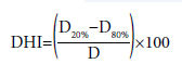
The dosimetric difference between IMRT and RA plans
was analyzed using the Statistical Package for the Social
Sciences (version 23; IBM Corp., Armonk, NY, USA)
in terms of the mean, standard deviation, and P-values.
The independent paired t-test with a confidence interval
limit of 95% was performed to assess the dosimetric
endpoints for the target and OARs. P-values of less
than 0.05 were used to denote statistical significance.
Clinically acceptable treatment plans were created using the RA and IMRT techniques for all patients. Qualitative and quantitative analyses were performed on dose distribution created for RA and IMRT plans. The data were derived from cumulative DVH data for each treatment plan.
Table
Figures
*: Signifies p?0.05. PTV: Planning Target Volume; IMRT:
Intensity-Modulated Radiotherapy; FF: Flattening filter; FFF:
Flattening filter-free.
*: Signifies p?0.05. IMRT: Intensity-Modulated Radiotherapy;
FF: Flattening filter; FFF: Flattening filter-free.
*: Signifies p?0.05. IMRT: Intensity-Modulated Radiotherapy;
FF: Flattening filter; FFF: Flattening filter-free.
*: Signifies p≤0.05. C: Coverage index; CI: Conformity index; HI: Homogeneity index; GI: Gradient
index; UDI: Unified dosimetry index; LGI: Low gradient index; HGI: High gradient index; IMRT:
Intensity-Modulated Radiotherapy; FF: Flattening filter; FFF: Flattening filter-free.
Figure
IMRT: Intensity-Modulated Radiotherapy; FF: Flattening filter; FFF: Flattening filter-free.
Figures
IMRT: Intensity-Modulated Radiotherapy; FF: Flattening filter; FFF: Flattening filter-free.
FF: Flattening filter; FFF: Flattening filter-free.
The DVH comparison between IMRT and RA plans
for FF and FFF plans is shown in Figures
PTV: Planning target volume; RTFH: Right femoral head; LTFH: Left femoral head; DVH: Dose-volume histogram; OARS: Organs at
risk; IMRT: Intensity-Modulated Radiotherapy; FF: Flattening filter; FFF: Flattening filter-free.
PTV: Planning target volume; RTFH: Right femoral head; LTFH: Left femoral head; RA: RapidArc; DVH: Dose-volume histogram;
OARS: Organs at risk; IMRT: Intensity-Modulated Radiotherapy; FF: Flattening filter; FFF: Flattening filter-free.
PTV Dose Distributions and Evaluations
As depicted in Figures 6, 7 of isodose distribution and
Figures 8, 9 of DVH, no difference in V95 dose distribution
was observed between IMRT FF and FFF plans. In
contrast, a significant difference was observed between
RA_FF and FFF plans, depicted in Figure 1. Moreover,
V98 showed a higher percentage of dose distribution in
plans with FF beams for both IMRT and RA planning
techniques, with a significant difference observed between
them (p<0.01).
Furthermore, there was a significant reduction in D50 observed in IMRT_FFF plans. Conversely, an increase in D50 was observed (p=0.18) with RA_FFF. Additionally, IMRT_FFF plans showed a decrease in Dmax (p=0.14), whereas RA_FFF plans demonstrated a significant increase in Dmax inside the PTV volume.
Plan Quality Indices
IMRT_FF and IMRT_FFF plans showed a homogeneous
plan with a 6 MV photon beam, but a significant
difference in homogeneity was observed with
RA_FFF plans. Furthermore, there was a significant difference of 14.26% (p<0.01) in DHI with RA_FFF
plans, whereas with IMRT_FFF, the difference was only 4.27%, which was insignificant. A highly conformal
plan was found with RA_FF and RA_FFF techniques. However, a significant difference was observed
with IMRT plans for FF and FFF.
The evaluation of UDI, LGI, and HGI indices revealed a significant difference in UDI between IMRT and RA plans utilizing the FFF beam. However, all the plans achieved similar plan quality indices in their respective techniques. Additionally, no differences were observed in LGI and HGI indices among plans using the FFF beam, IMRT, and RA.
Dose Sparing of the OARs
Bladder: There was a significant increase in V30, V40,
and Mean Dose (Dmean) observed with the IMRT_FFF
beam; however, a reduction in V45 was found with
IMRT_FFF (p<0.05). Furthermore, no significant difference
was observed in the near-max dose (D2cc). For
RA_FFF, a decrease in V45 was found (p=0.07), and a
significant increase in D2cc was observed (p<0.01).
Rectum: A significant increase in V30, V40, and Mean Dose (Dmean) was found with IMRT_FFF plans. However, no difference was observed in V45 and D2cc. Furthermore, no significant difference was found between RA_FF and RA_FFF plans; a decrease in V45 was found with RA_FFF plans.
A large percentage reduction in V45 was observed with IMRT plans compared to RA for both the bladder and rectum, as shown in Figures 2, 3.
Bowel: The RA_FFF plans showed a reduction in V40, but no improvement was observed in V45. On the contrary, there was a significant increase in V40 with the IMRT_FFF technique; however, a significant reduction was observed in V45. Moreover, a similar scenario was observed with RA_FFF, but these differences are not statistically significant.
RTFH & LTFH: In both femoral heads, no significant differences were found between IMRT_FF and IMRT_ FFF, except in V30 (p<0.05). However, a significant increase in femoral head dose was found with RA_FFF plans.
The total number of monitor units (MU) is important for assessing the low dose to normal tissue and treatment time. The present study observed a significant increase in monitor units with FFF beam plans for IMRT and RA techniques. Moreover, the percentage difference in the increase in MUs is less in RA (12.16%) plans compared to IMRT (32.46%) plans.
The quantitative analysis of Figure 5 showed that dose fall-off beyond the target region was similar for all the datasets. Furthermore, we have taken ln(D) vs. ln(1/V) to evaluate the rate of dose fall-off beyond PTV. The fall-off shows that for low dose volumes (V1, V2, V3, V4, and V5), the change in dose fall-off is similar, which continues till V10. However, a steep dose fall-off was observed with intermediate dose volumes (V20, V30 and V40), starting from V20, which clearly shows a steeper dose gradient found with RA plans with FF compared to FFF for V20 and V30 (p<0.01).
FFF beam plans showed similar target coverage and
increased bladder, rectum, and femoral head protection
with RA techniques compared to IMRT. Furthermore,
RA plans achieved less MU than IMRT. The RA_FFF
plans showed a higher maximum dose to PTV, as presented
by Dmax and D2, compared with IMRT_FFF for
better target coverage. Furthermore, with both techniques,
a significant reduction in V98 was found with
the FFF beam, as shown in Figure 3. Similar results were
found by Manna et al.[
In the current study, we have used many plan quality
indices to qualitatively analyze the treatment plans
generated by the FFF beam compared to FF for IMRT
and RA techniques. The results are compared to find out
the effective treatment plan for the treatment of postop
cervical cancer patients. Our study showed that RA
produces more conformal and homogeneous plans than
IMRT. This was consistent with the report that more
conformal and homogeneous plans using the RA technique
for post-operative cervical cancer patients.[
However, there was no difference in the Gradient
Index for RA and IMRT plans. With FFF, most of the
quality indices showed non-significant variation, except
CI with IMRT_FFF plans and HI with RA_FFF plans. In addition, a decrease in UDI value was observed
with IMRT_FFF, as shown in Figure 4, although this
is not statistically significant. Zhang et al.[
For OAR, the doses are smaller for FF than for FFF;
partly, this can be traced to a smaller part of the PTV
receiving the minimum dose required by the objectives.
FFF for both bladder and rectum showed a decrease
in dose for V45; however, this is not statistically
significant. Furthermore, an increase in dose in dosimetric
parameters (V30, V40) was found with the FFF
beam for both the IMRT and RA techniques. However,
these differences are small too, in most cases less than
1% of the volume of the OAR, especially in comparison
to the large standard deviation. The previous study
with IMRT and RA showed a minimal improvement in
dose to OARs with the FFF beam.[
Further, after a hysterectomy, the small bowel falls
into the pelvis where the uterus previously resided, further
increasing the amount of small bowel irradiated
to the prescription dose. Rates of grade 2 and higher
acute gastrointestinal (GI) toxicity of 50-90% with
conventional CRT have been reported in the literature.
Acute GI symptoms typically involve varying degrees
of diarrhea, cramping, and abdominal pain, which can
negatively impact the quality of life during treatment.
[
Previous studies on the IMRT technique used a
PRO optimizer to generate their FF and FFF beam
plans.[
There was a significant increase in MU observed
with the FFF beam for IMRT (32.5%, p<0.01) and RA
(12.2%, p<0.01) techniques. Though the delivery time
of the FFF beam (Dose rate 1440 MU/min) is higher
than FF (600 MU/min), the increase in MU is related to
achieving uniform dose distribution with an inhomogeneous
profile. This finding is in line with the results
from previous studies.[
Ethics Committee Approval: The study was approved by the Institutional Ethics Committee (no: IEC/11/65/2023, date: 12/07/2023).
Authorship contributions: Concept - S.M., S.S., P.K.G., R.T.; Design - S.M., S.S., P.K.G., R.T.; Supervision - S.S., P.K.G.; Data collection and/or processing - S.M., R.T.; Data analysis and/or interpretation - S.M., S.S., P.K.G., R.T.; Literature search - S.M., S.S., P.K.G.; Writing - S.M., S.S., P.K.G.; Critical review - S.M., S.S., P.K.G., R.T.
Conflict of Interest: All authors declared no conflict of interest. Use of AI for Writing Assistance: No writing assistance was utilized in the production of this manuscript.
Financial Support: This research did not receive any specific grant from funding agencies in the public, commercial, or not-for-profit sectors.
Peer-review: Externally peer-reviewed.

